Notes from a California naturalist

The nature of Nature

Menu
  • About me
  • Contact me
Menu

Tag: natural history

Blitzin’ the intertidal, part 2

Posted on 2017-06-292023-01-06 by Allison J. Gong

The intertidal portion of my participation in Snapshot Cal Coast 2017 is complete. I organized four Bioblitzes, two of which consisted of myself and Brenna and the other two for docents of the Seymour Marine Discovery Center (Tuesday) and the docents of Año Nuevo and Pigeon Point State Parks (Wednesday). The four consecutive days of early morning low tides have been exhausting for a concussed brain and a body dealing with bronchitis for the past several weeks. Good thing the low tide arrives 40-50 minutes later, or I’d probably be dead by now. And even so, I tried to take advantage of the later tides to venture a bit farther afield, so I still ended up getting up at the butt-crack of dawn.

But oh, so totally worth it!

Day 3: Davenport Landing with docents from the Seymour Marine Discovery Center, Tuesday 27 June 2017, low tide -1.1 ft at 08:03

Davenport Landing Beach is a sandy beach with rock outcrops and a fair amount of vertical terrain to the north, and a series of flat benches (similar to those at Natural Bridges) to the south. To get to the good spots at the north end you have to do some cliff scrambling, unless the tide is low enough that you can walk around the rock, which happens maybe once or twice a year. Because it’s easier to get around on the benches to the south, that’s where I took my group for the Bioblitz. The difference in topography also results in some differences in biota and distribution/abundance of organisms; overall biodiversity is probably equivalent at both sites, but certain species are more abundant at one site versus the other.

Intrepid citizen scientists at Davenport Landing
27 June 2017
© Allison J. Gong

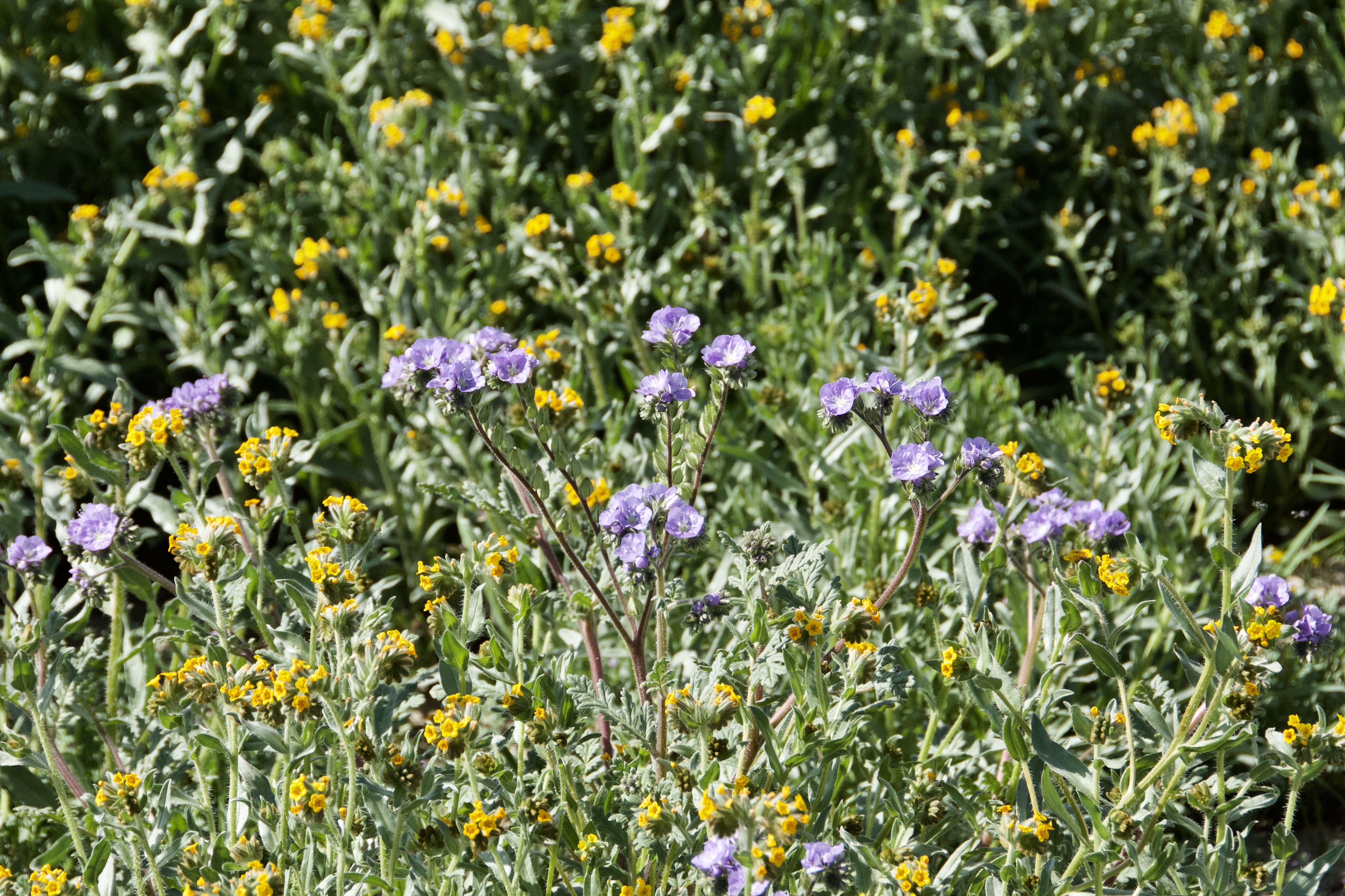
The morning we went to Davenport was sunny and (almost) warm. This makes for plenty of light for photography, but also lots of glare of the surface of pools and the wet surfaces of organisms themselves. My most successful photos are the ones I took with the camera underwater. Wanting to improve my skills at identifying algae, I concentrated most of my efforts on them while not ignoring my beloved invertebrates.

Encrusting coralline algae on submerged rock
27 June 2017
© Allison J. Gong

Coralline algae are red algae whose cells are impregnated with CaCO3. This gives them a crunch texture that is unusual for algae. Corallines come in two forms, encrusting and upright, and can be one of the most abundant organisms in the high and mid intertidal. There are several species of both encrusting and upright corallines on our coast, and most of the time they aren’t identifiable to species by the naked eye. Sometimes I can distinguish between genera for the upright branching species. However, the encrusting species require microscopic examination of cell size, crust thickness, and reproductive structures, none of which can be observed in the field.

Bullwhip kelp, Nereocystis luetkeana, at Davenport Landing.
27 June 2017
© Allison J. Gong

Some algae are so distinctive that a quick glance is all it takes to know exactly who they are. With its tiny holdfast, long elastic stipe, and single large pneumatocyst, bullwhip kelp doesn’t look anything like the other kelps in California. Like most kelps, N. luetkeana lives mostly in the very low intertidal or subtidal, where under certain conditions it can be a canopy-forming kelp. About a month ago I noted a big recruitment of baby Nereocystis kelps in the intertidal on the north side of Davenport Landing Beach. I speculated then that they probably wouldn’t persist into the summer. I’ll have to take a morning soon to go up and check on them. Anyway, on our Tuesday Bioblitz we found this big N. luetkeana growing in the intertidal. The stipe was about 1.5 meters long and the pneumatocyst was a little smaller than my closed fist. Given that this individual recruited to that spot and has persisted for a few months, probably, it has a good chance of continuing to survive into the fall. Winter storms, especially if they’re anything like the ones we had this past year, will most likely tear it off, though.

Coralline algae aren’t the only pink things in tidepools. There are pink fish!

Sculpin in tidepool at Davenport Landing.
27 June 2017
© Allison J. Gong

Sculpins are notoriously difficult to ID if you don’t have the animal in hand to count things like fin rays and spines. Someone on iNaturalist may be able to ID this fish, but I don’t think the photo is very helpful.

And, just because they’re my favorite photographic subjects in the intertidal, here’s a shot of Anthopleura sola:

Anthopleura sola at Davenport Landing
27 June 2017
© Allison J. Gong

As of this writing, 10 participants in this Bioblitz have submitted 204 observations to iNaturalist, with 70 species identified. I know that some people haven’t upload their observations yet, and expect more to come in the next couple of weeks. The docents enjoyed themselves, to the extent that two of them accompanied Brenna and me to our fourth Bioblitz at Pigeon Point.


Day 4: Whaler’s Cove at Pigeon Point with rangers (and one docent) from Pigeon Point and Año Nuevo state parks, Wednesday 28 June 2017, low tide -0.6 ft at 08:53

Usually when I go to Pigeon Point I go to the north side of the point, either scrambling down the cliff next to the lighthouse or about half a mile north to Pistachio Beach. When the park rangers and I were organizing this Bioblitz they suggested going to Whaler’s Cove, as the access is very easy due to a staircase and would be much easier for docents who aren’t used to climbing down cliffs. It ended up being a good decision, as there was much to be seen.

Bioblitzes and iNaturalist are all about photographing individual organisms (as much as possible) so that they can be ID’d by experts in particular fields. This is the ‘tree’ level of observation I mentioned in my previous post. I find that when I’m taking photos with the intent to upload them to iNaturalist the photos themselves tend to be rather boring. The intertidal is such a dynamic and complex habitat that photos of single species tend to lack the visual interest of the real thing. I’ve learned that one of my favorite things to see is organisms living on other organisms.

See what I mean?

A nicely decorated mossy chiton, Mopalia muscosa, at Pigeon Point.
28 June 2017
© Allison J. Gong

Four of this chiton’s eight shell plates are completely covered with encrusting coralline algae. It is also wearing some upright corallines and at least two other red algae, one of which is Mastocarpus papillatus. This photo produced six observations for iNaturalist.

Which is not to say that single-subject photos are always boring. When the subject is as weighty as this gumboot chiton (Cryptochiton stelleri), it deserves its own photo or two.

Cryptochiton stelleri at Pigeon Point
28 June 2017
© Allison J. Gong
Ventral view of Cryptochiton stelleri
28 June 2017
© Allison J. Gong

The largest chiton in the world, Cryptochiton typically lives in the subtidal or the very low intertidal. Unlike other chitons, it doesn’t stick very firmly to the substrate. I was able to reach down and pick up this one with very little effort. In the subtidal this lack of suction isn’t a handicap, as water movement there is less energetic compared to the intertidal, and Cryptochiton does quite well. But it doesn’t really look like a chiton at all, does it? That’s because its eight dorsal shell plates are covered by a thick, tough layer of skin called the mantle. In most chiton species the mantle is restricted to the lateral edges of the dorsal surface. The girdle, as it’s called, exposes the shell plates to some degree. We don’t see Cryptochiton‘s shell plates, but if you run your finger down the middle of the dorsum you can sort of feel them underneath the mantle.

Okay, now for some more ‘forest’ pictures.

Intertidal biota at Pigeon Point
28 June 2017
© Allison J. Gong

I love this one. There’s a lot going on in this small area. The greenish-brown algae are actually a red alga, Mazzaella flaccida. There are two large clumps of stuff in the photo. The clump on the left, consisting of round lumps, is a clone of the aggregating anemone Anthopleura elegantissima. The other clump is a mass of tubes of the polychaete worm Phragmatopoma californica. These two clumps were formed in very different ways, reflecting the vastly different biology of the animals that made them.

Anthopleura elegantissima is one of four species of Anthopleura anemones we have in California and is the only one to grow by cloning. It does so via longitudinal fission, in which an anemone literally rips itself in half. I wrote about them last year. Note that in this aggregation, all of the anemones are about the same size. That’s because they’re all clones of each other and share the exact same genetic makeup.

Whereas a clone of A. elegantissima represents a single genotype formed by cloning, clumps of Phragmatopoma arise by gregarious settlement. Each of the tubes in a clump is occupied by a single worm, which recruited to that spot as a larva and settled down to live its life. When it comes time to look for a permanent home, the planktonic larvae of Phragmatopoma are attracted by the scent of adult conspecifics. The larvae settle on the tubes of existing adults and undergo metamorphosis. Each worm builds its tube as it grows, using some kind of miraculous cement that sticks sand grains together, much as a mason stacks bricks to build a wall. One of the remarkable things about this construction is that the cement is secreted by the animal’s body and starts out sticky and then hardens, all in seawater. It’s a likely candidate for Best Underwater Epoxy around. Interestingly, Phragmatopoma can build its tube only as a growing juvenile. Adult worms that are removed from their tubes do not build new ones, and soon die.

Here’s another nice clump of Phragmatopoma:

Intertidal biota at Pigeon Point
28 June 2017
© Allison J. Gong
Whaler’s Cove at Pigeon Point
28 June 2017
© Allison J. Gong

See that pile of rocks out there? That’s where we were blitzing. Given the not-so-lowness of the tide I didn’t know if we would be able to make it out there. We were lucky, though, and were able to spend ~30 minutes out on that little point.

So far, the Pigeon Point Bioblitz has yielded 204 observations for iNaturalist, with three participants (so far!) identifying 77 species. Several of my observations were of red algae that I did not recognize; hopefully an expert will come along to ID those for me. Snapshot Cal Coast 2017 continues through this weekend. My intertidal Bioblitzes are over, but I hope to contribute one last set of observations by collecting and examining plankton on Sunday.

Share this:

  • Share on Reddit (Opens in new window) Reddit
  • Email a link to a friend (Opens in new window) Email
  • Share on Mastodon (Opens in new window) Mastodon
  • More
  • Share on Tumblr

Like this:

Like Loading...

Blitzin’ the intertidal, part 1

Posted on 2017-06-262023-01-06 by Allison J. Gong

This is the second year that the California Academy of Sciences has sponsored Snapshot Cal Coast, a major effort to document and characterize the biodiversity of the California coast. To this end the Academy has organized several Bioblitzes at various sites in northern California, and solicited volunteers to lead their own Blitzes, either as individuals or with groups. A Bioblitz is a citizen science activity in which people take photographs of organisms or traces of organisms (shells, scat, tracks, etc.), then upload their observations into iNaturalist. Experts then identify the organisms in the observations, and the data are publicly available to anyone who wants to use them.

For Snapshot Cal Coast 2017 I have four Bioblitzes planned for the intertidal. Here are some of my observations made in the first two.

Day 1: Natural Bridges, Sunday 25 June 2017, low tide -1.7 ft at 06:27

My friend Brenna joined me on an early low tide at Natural Bridges. The intertidal topography at Natural Bridges consists of a series of gently sloping benches that are riddled with potholes of various sizes and depths. For the purposes of this Bioblitz I decided to confine my observations to the geological structure that I call the peninsula, which sticks out farther into the ocean than the edges of the benches.

Aerial view of intertidal benches at Natural Bridges, with the “peninsula” circled in red.
26 June 2017
© Google Maps

The peninsula is most easily accessible when the tide is at least as low as -1 ft, although large swell can make it entirely unsafe to do so at even very low tides. Fortunately the swell wasn’t big enough to keep me from the peninsula yesterday, and I confined most of my observations to this location. I’ve found that making observations for Bioblitzes requires a different kind of attention and focus than either collecting or observing for more general purposes. In the spectrum of forest-to-trees levels of observation, Bioblitzes are all about individual trees. When left to my own devices I tend to move quite fluidly between forest-level observations (e.g., broadscale ecological patterns) and tree-level observations (e.g., what organism is that?), and confining myself to only tree-level observations was, well, confining. It’s undoubtedly a good discipline, but one that I find a little stifling.

Here are some of the “trees” I saw at Natural Bridges.

Anthopleura sola
25 June 2017
© Allison J. Gong
Haliotis cracherodii, the black abalone
25 June 2017
© Allison J. Gong

I’ve been keeping an eye on this abalone for a couple of years now. It has gotten bigger and in the last year has become heavily encrusted with other animals and algae. Right now it is sporting lots of acorn barnacles (both large and small), at least one tube of Phragmatopoma californica, limpets, encrusting and upright coralline algae, and other red algae.

The red alga Smithora naiadum 
25 June 2017
© Allison J. Gong

Smithora naiadum is a red alga whose thallus consists of small flat blades. It grows only as an epiphyte on seagrasses, in this case the surfgrass Phyllospadix scouleri. Later in the summer many surfgrass leaves will be almost entirely covered with Smithora.

My favorite observation of the morning was this little hermit crab.

Pagurus hirsutiusculus, the so-called hairy hermit crab
25 June 2017
© Allison J. Gong

I love how this hermit is clinging to a piece of giant kelp. It lives in a shell of the olive snail Olivella biplicata, as many of its conspecifics do. These shells get to a bit over 2 cm in length, and their narrow diameter means there isn’t much empty space inside. Fortunately, P. hirsutiusculus is one of the smaller hermit crabs and doesn’t need much space.

An extreme low tide like yesterday’s has two benefits. The most obvious is that more real estate is exposed, thus more area to explore. The second benefit of a really low tide is time. Much of the biodiversity of the intertidal is in the low-mid and low zones; the lower the tide, the longer it takes for the ocean to return and reclaim its property. I was able to spend the better part of two hours out on the peninsula, which doesn’t happen every year. Lucky me!


Day 2: Franklin Point, Monday 26 June 2017, low tide -1.5 ft at 07:15

To get to the beach at Franklin Point you have to hike ~10 minutes over the dunes along a maintained trail. The views along the way are often quite spectacular, even when it’s foggy. This morning it was unusually clear, and I wished I had brought along my big camera. For example, looking north towards Pigeon Point I saw this:

View towards Pigeon Point from the Franklin Point trail
26 June 2017
© Allison J. Gong

I mean, come on. How much more beautiful can a vista be?

The intertidal at Franklin Point has changed dramatically over the past year. Heavy storms over the 2016-2017 winter removed about two vertical meters of sand from the beach, exposing rocks that had been buried for years. Even today, months after the peak of the storm season, you can see bare rock that has yet to be heavily colonized by living things.

Mostly bare rock at Franklin Point
26 June 2017
© Allison J. Gong

Primary succession is the sequence of species’ arrival and eventual replacement in an area that has never hosted life before. These rocks may very well have served as habitat for organisms years ago, but in my memory they had been buried in sand until the recent storms. Their exposure provides an opportunity to observe primary succession in this very dynamic habitat.

The first organisms to arrive and take hold in any newly available habitat are primary producers. Makes sense, as there is no food for heterotrophs yet. In the case of the intertidal the first visible organisms are algae. The algae at Franklin Point have been going like gangbusters all spring and into the summer. Faunal diversity, on the other hand, has been rather low. I spent quite a while looking at and photographing algae, many of which I couldn’t identify in the field.

My favorite red alga, Erythrophyllum delesserioides, at Franklin Point
26 June 2017
© Allison J. Gong
A young specimen of Egregia menziesii at Franklin Point
26 June 2017
© Allison J. Gong

Some things were entirely unfamiliar to me. For example, I’d never seen coralline algae encrusting on the tips of another red alga. And yet, here it is:

Coralline algae
26 June 2017
© Allison J. Gong

As I mentioned above, animal life at Franklin Point has been rather depauperate this year. HOWEVER, I did get to let out a few whoops of triumph when I found this:

The staurozoan Haliclystus sp. at Franklin Point
26 June 2017
© Allison J. Gong

These animals, staurozoans, are incredibly difficult to photograph. Not only are they the same color as many of the algae they live with and attach to, but they like areas where the water is constantly moving back and forth. Plus, the pools and channels where I found them were cloudy with Ulva spooge. I took a lot of pictures of backscatter and blurry staurozoans.

Here’s another shot:

Haliclystus sp. at Franklin Point
26 June 2017
© Allison J. Gong

Staurozoans are the strangest and by far the coolest cnidarians. Their common name ‘stalked jellyfish’ harkens back to when they were considered scyphozoans, close kin to moon jellies (Aurelia) and the like. They are now known to be in their own group, the Staurozoa, related to but not part of the Scyphozoa.

I don’t really know why I’m so enamored of the staurozoans. Maybe it’s because they are rare and poorly understood. I know them only from Franklin Point and one sighting at Carmel Point. The systematics of the staurozoans is in flux; I’m not brave enough to assign a species epithet to this critter, but a colleague who is one of the people working on this group suggests that it is H. sanjuanensis, a species that has not yet been formally described. All of the staurozoans I saw today were this brownish-red color, but in previous years I’ve also seen them in a brilliant bottle green. Those would probably be easier to see among all the red algae, but with my luck the green ones would all be hanging out with Ulva.

The very last part of the hike to the intertidal is a steep decline down the dune to the beach. Getting down is easy, you just sort of ski down. Getting up is much more of a challenge. Ever try to climb a sand dune? Each step gets you about a quarter of a step above the last one, so it’s hard work, especially when the dune is steep. There have been times that I’ve hiked all the way out to the beach, only to turn around and go back because I didn’t think I’d be able to climb back up the dune in my hip boots. And since I have bronchitis right now by the time I got back to the top today it felt as though I had climbed Mt. Everest.

See?

It’s steeper than it looks, especially on the way up
26 June 2017
© Allison J. Gong

All told, I added about 150 observations to iNaturalist these first two Bioblitzes. I’m not really into making observations just to make observations, so for me that 150 is a good two days’ production. Now I need to rest up for tomorrow’s low tide.

Share this:

  • Share on Reddit (Opens in new window) Reddit
  • Email a link to a friend (Opens in new window) Email
  • Share on Mastodon (Opens in new window) Mastodon
  • More
  • Share on Tumblr

Like this:

Like Loading...

Emblem of the Golden State

Posted on 2017-05-252023-01-06 by Allison J. Gong

Did you know that California has a state lichen? I didn’t either, and it turns out that we’ve had one for over a year! In January of 2016, California became the first state to adopt an official state lichen, and Ramalina menziesii joined the ranks of the California poppy (Eschscholzia californica), the California quail (Callipepla californica), the coast redwood (Sequoia sempervirens), and the extinct-in-the-wild California grizzly bear (Ursus californicus) as official symbols of the Golden State.

The lichen Ramalina menziesii growing on a coast live oak (Quercus agrifolia) at Rancho del Oso
29 January 2016
© Allison J. Gong

Lichens are strange beasts, resulting from a symbiosis between two very different organisms, an alga (or in some cases a cyanobacterium) and a fungus. They are photosynthetic like plants and algae thanks to the algal/cyanobacterial partner in the symbiosis, but do not have roots or leaves. The fungus component restricts them to places where fungi can live, which means you generally don’t find lichens in very dry places. That said, some lichens have adapted to live in hostile habitats such as the Arctic tundra and arid deserts. Many of them live on trees and other plants, but when they do so they are not parasitic. They can grow on nonliving surfaces such as rocks, buildings, and soils. Lichens are crucial players in the ecological process of primary succession, which occurs when virgin habitat is newly opened up to colonization by life (for example, the area left scoured by a retreating glacier, or land formed by recent lava flowing into the sea). The fungal partner of a lichen sends out hyphae which burrow into rock, eventually weakening it and forming soil. Plants cannot take root until soil is present, so lichens, in addition to being among the first organisms to colonize an area, modify the habitat to enable other species to become established.

In some ways, the fungus partner of a lichen can be viewed as a farmer, in the sense that it houses photosynthetic symbionts that do the hard work of fixing carbon into molecules such as sugars, which can then be used to fuel the fungus’s metabolism. The fungus doesn’t just mooch off its symbionts, though. As in other symbiotic relationships between unicellular algae and multicellular hosts, the fungus provides a safe place for the algae to live, as well as a stable environment in which to carry out its photosynthetic magic. 

Ramalina menziesii at Rancho del Oso
29 January 2016
© Allison J. Gong

Most lichens have a simple morphology, growing as a crust over the substrate. Ramalina menziesii has a lacy morphology and typically lives as an epiphyte, draping over the branches of trees and shrubs. It is often associated with oak trees in California, especially the Coast Live Oaks (Quercus agrifolia) that live in the more humid regions along the coast. During the drought there was much less Ramalina hanging from the thirsty oak trees, but this year there does seem to be more of it. Strands of R. menziesii are used as nesting material by many birds, and I’ve seen deer eating whole gobs of the stuff, pulling it off the trees with their rubbery lips.

Lichens, including Ramalina menziesii, growing on a Coast Live Oak (Quercus agrifolia) at Los Osos Oaks Reserve in San Luis Obispo County
2 January 2015
© Allison J. Gong

Ramalina menziesii is often referred to as “Spanish moss” which is misleading on any number of counts. First of all, it’s not Spanish, being a species native to the west coast of North America. Second, it’s not a moss; mosses are plants, and Ramalina is a lichen. Third, there is a true flowering plant (a bromeliad, actually, not a moss at all) with the common name Spanish moss that lives as an epiphyte in the warm humid southeastern U.S. as well as other tropical areas; clearly, this is not the same organism as R. menziesii, although the two may share superficial similarities such as overall growth form and color. If R. menziesii requires a common name for people to understand what it is, then let that name be something descriptive and biologically accurate, such as “lace lichen”; I’ve seen this name on a few websites and like it.

Lichens and fungi comprise a large part of my body of ignorance regarding the natural history of California. I find them very interesting but inscrutable, and they don’t speak to me as loudly as do my beloved marine invertebrates. What this means is that I have a lot of learning to do, and this is always a Good Thing.

Share this:

  • Share on Reddit (Opens in new window) Reddit
  • Email a link to a friend (Opens in new window) Email
  • Share on Mastodon (Opens in new window) Mastodon
  • More
  • Share on Tumblr

Like this:

Like Loading...

Peanut worms!

Posted on 2017-05-232023-01-06 by Allison J. Gong

If I ask my invertebrate zoology students to name three characteristics of the Phylum Annelida, they would dutifully include segmentation and chaetae (bristles) in the list. And they would be correct. Annelids, for the most part, are segmented and many of them have chaetae. But in biology there are many exceptions for every rule we teach, and it’s these exceptions that make a deeper study of biology so rewarding.

Peanut worms (Phascolosoma agassizii) at Pigeon Point
30 April 2017
© Allison J. Gong

A couple of weeks ago I did some collecting in the intertidal at Pigeon Point. It was a very accommodating low tide, and I had a lot of time to poke around and explore. I found an area that had several decently sized rocks that I could turn over, and had fun seeing what lives on the side away from the light. Some of the animals on the underside of rocks are the common ones you see everywhere–turban snails, limpets, Leptasterias stars, and the like. Some, however, prefer a life of darkness and actively move away from the sun when their rock is turned over. And others happen to live in the sand under the rock and might not care one way or the other about the light.

Peanut worms, scientifically known as sipunculans, are delightful small worms that in my opinion are vastly underappreciated. This is understandable, as they are usually hidden in sand or rubble and aren’t exactly conspicuous even when uncovered. Phascolosoma agassizii is our local sipunculan. Like all sipunculans it is unsegmented, and it has no chaetae. Peanut worms used to be elevated to their own phylum, the Phylum Sipuncula; however, molecular evidence shows that they are indeed annelids despite their apparent loss of key features such as body segmentation and chaetae.

Peanut worms (Phascolosoma agassizii) at Pigeon Point
30 April 2017
© Allison J. Gong

They do look vaguely peanut-ish, don’t they? They’re small, maybe 6 cm all stretched out, which you hardly ever see. Phascolosoma agassizii is a grayish pink color, with irregular black stripes that usually don’t form complete hoops around the body. Peanut worms are sedentary, living with most of the body buried in sand, rubble, shell debris, kelp holdfasts, etc. One of the weird things about them is that the mouth in located on the distal end of a long tube called the introvert. Most of the time the introvert is stuffed inside the main body region, or trunk. It is eversible and unrolls from the inside out, sort of like when you remove a long sock by pulling the top edge down over your leg and off your foot. The mouth on the end of the introvert is surrounded by short sticky tentacles, and the introvert dabs around to pick up organic deposits from the surfaces. Mucus and cilia on the tentacles convey the yummy organic gunk to the mouth, and a pharynx pushes food through to a long esophagus that runs the length of the introvert and leads to the long coiled intestine in the trunk.

Watch these peanut worms extending and retracting their introverts. Cute, aren’t they?

I brought three peanut worms back to the lab with me, where they are happily living in my sand tank. Their housemates are ~15 sand crabs (Emerita analoga) and a clump of tube-dwelling polychaetes (Phragmatopoma californica). I never see them unless I dig them up from the sand, which leads me to believe that they do most of their feeding at night. Either that or they actually do actively shy away from the light.

Despite not sharing much in the way of apparent morphological similarity with more typical annelids, sipunculans are indeed annelid-like in other ways. Many of their internal structures are like those of annelids, and at least their early development (cleavage pattern and differentiation of tissue layers) follows the annelidan pathway. The species that have indirect development have a trochophore larva, typical of the marine annelids, that in some cases morphs into a second larval stage called a pelagosphera.

Sipunculans are the poster child for Animals That Are Not What They Seem. But they are interesting in their own way, and I always have a “yay!” moment when I find them in the field. It’s really hard not to make sound effects as they’re rolling their introverts in and out. You should try it yourself some time.

Share this:

  • Share on Reddit (Opens in new window) Reddit
  • Email a link to a friend (Opens in new window) Email
  • Share on Mastodon (Opens in new window) Mastodon
  • More
  • Share on Tumblr

Like this:

Like Loading...

Eight is enough

Posted on 2017-05-162023-01-06 by Allison J. Gong

One of the defining characteristics of the Phylum Mollusca is the possession of a shell, which serves both as a protective covering and an exoskeleton. We’ve all seen snails, and some people may have noticed that snails often withdraw entirely into their shells and even have a little door that they can use to seal up the opening of the shell. That little door is called the operculum. Opercula occur in non-molluscan animals, too, such as some of the tube-dwelling polychaete worms and some of the thecate hydroids. Snail opercula come in lots of different shapes, depending on the aperture of their owner’s shell.

Calliostoma ligatum at Mitchell’s Cove
2 April 2017
© Allison J. Gong

Given the enormous morphological diversity within the Mollusca it shouldn’t be surprising that their shells vary immensely in prominence and shape. In fact, molluscan shells demonstrate quite beautifully the relationship between form and function. The benthic and most familiar molluscs, the gastropod snails, generally have coiled shells. Notable exceptions to this generality are the marine opisthobranchs (nudibranchs and sea hares) and the terrestrial slugs. And for the most part snail shells look recognizably like snail shells, even though some are plain coils, others may be flattened (e.g., abalones), and still others may be crazily ornamented. Aquatic animals crawl around in water, which helps to support the weight of heavily calcified shells. Terrestrial snails, on the other hand, live in a much less dense medium (air) and have lighter, less calcified shells. The trade-off for a more easily transportable shell is that air is also very drying, and a thinner shell provides less protection from desiccation.

I should state for the record right now that I’m not talking about the many molluscs that don’t have shells at all, or that have much reduced shells.

View into the exhalant opening of a mussel (Mytilus sp.) at the Santa Cruz Yacht Harbor
29 August 2015
© Allison J. Gong

The bivalve molluscs (mussels, clams, oysters, etc.) live inside a pair of shells. They are sedentary animals, living either attached to a hard surface or buried in sand or mud. Not being able to run from predators (although some scallops can swim!), their only defense is the toughness of their shells and the strength of the adductor muscles that hold the shells closed. Most bivalves feed by sucking water into the shells through an incurrent siphon, using their gills to filter food particles from the water, and expelling the water through an excurrent siphon. To do so they must open their shells enough to extend their siphons, or at least expose inhalant and exhalant openings, to the water current surrounding them.

So, snails have one shell and bivalves have two. Some of the most interesting molluscs, in terms of shell morphology, are the chitons. The Polyplacophora (Gk: ‘many plate bearer’) have a shell that is divided into eight dorsal plates. This makes them immediately distinguishable from just about any other animal.

Chiton (Lepidozona mertensii) at Point Piños
6 February 2016
© Allison J. Gong

Chitons live from shallow water to the deep sea, but the majority of species live in the intertidal. This is a high-energy habit characterized by the bashing of waves as the tide rises and falls twice daily. Any organism living here must be able to hang on for dear life or risk being swept away to certain death. Chitons are certainly well equipped to survive in this habitat. They have a low profile, offering minimal resistance to the waves. Rather than stand tall and face the brunt of the wave energy, chitons cling tightly to the rocks and let the waves wash over them.

Tonicella lokii at Monastery Beach
27 October 2015
© Allison J. Gong

The chiton’s shell, divided into eight articulating plates, gives the animal a much more flexible shell than is found in any other mollusc. This allows them to conform to the topography of the rocks, giving them an even lower profile than, say, a limpet of the same overall shape and size.

While most chitons are pretty sedentary, at least during the low tides when we can see them, some of them can move pretty quickly when they want. So what, exactly, motivates a chiton to run? One species, Stenoplax heathiana, lives on the underside of rocks in the intertidal; it comes out at night to forage on algal films and retreats back under its rock with the dawn. I’ve seen them at Pistachio Beach, where I turned over rocks and watched them run away from the light. This video is shot in real-time; the chitons are really running fast!

When the eight shell plates are visible it’s easy to identify a chiton as a chiton. But not all chitons are quite so obliging with their most chiton-ish characteristic, and one is downright misleading.

Below is Katharina tunicata, one of the largest chitons on our coast. Its shell plates are barely visible, as they are almost entirely covered by the animal’s mantle, the layer of tissue that covers the visceral mass and encloses an open space called the mantle cavity in which the gills are located. In chitons, the mantle extends onto the dorsal side of the animal and is called the girdle. Katharina‘s girdle is smooth and feels like wet leather.

The chiton Katharina tunicata at the Great Tidepool in Pacific Grove
26 October 2015
© Allison J. Gong

The largest chiton in the world is the gumboot chiton, Cryptochiton stelleri, and it lives on our coast. This beast is about the size of a football, reaching a length of 30 cm or so. It lives mostly in subtidal kelp forests, but can be found in the very low intertidal, which is where I usually see it. At first encounter it’s hard to figure out what this animal is. It certainly doesn’t look like a chiton.

A large Cryptochiton stelleri at Mitchell’s Cove
6 June 2016
© Allison J. Gong

If anything, it looks like a mostly deflated football, doesn’t it? Turning it over to look at the underside doesn’t help much, either, although this photo does give an idea of how big the animal can get:

Ventral view of Cryptochiton stelleri at Pigeon Point
24 April 2016
© Allison J. Gong

Cryptochiton goes one beyond Katharina and covers its plates entirely. Just looking at the animal you’d have no idea that there are eight plates underneath the tough reddish-brown mantle, but you can feel them if you run your finger along the midline of the dorsum. Living subtidally as it does, Cryptochiton doesn’t have the ability to cling tightly to rocks that its intertidal relatives do, and it tends to get washed off its substrate and cast onto the beach during storms. I’ve never seen one on the beach that wasn’t very dead. Once a friend and I were trudging back up the beach after working a low tide, and encountered a dead softball-sized Cryptochiton. I mentioned that it would be nice to have a complete set of shell plates from one of these animals. My friend always carries a knife in her pocket, so we started an impromptu dissection right there on the beach. It didn’t take long to learn that the mantle of a gumboot chiton is really tough and difficult to cut through with a pocket knife. And even once we got through the mantle, dissecting the plates from the underlying tissue wasn’t going to happen with the tools we had with us. Besides, the stench was godawful even with our unusual tolerance for the smell of dead sea things. We abandoned that corpse.

Single plate of the gumboot chiton, Cryptochiton stelleri
16 May 2017
© Allison J. Gong

Many beachcombers have found white butterfly-shaped objects in the sand, but not known what they are. They are definitely calcareous and feel like bone, but what kind of animal makes a bone shaped like this? Turns out this object is one of the shell plates from C. stelleri. They wash up frequently, never attached to their neighbors so they provide no clue as to what organism they came from.

In order to obtain a complete set of Cryptochiton plates, I’d have to start with an intact chiton corpse. I did happen upon another dead Cryptochiton on a beach somewhere I was allowed to collect organisms, and I brought it back to the marine lab. I remember spending a smelly afternoon cutting the plates out of the corpse and removing as much of the tissue as I could, then feeding the plates to various hungry anemones to take care of the rest. Some of the plates got a little broken during the extraction process, but I do have my very own full set!

Shell plates of Cryptochiton stelleri
16 May 2017
© Allison J. Gong

Some day I will figure out a way to mount those plates permanently.

One final question to ponder. Does a chiton have one shell, or eight shells?

Share this:

  • Share on Reddit (Opens in new window) Reddit
  • Email a link to a friend (Opens in new window) Email
  • Share on Mastodon (Opens in new window) Mastodon
  • More
  • Share on Tumblr

Like this:

Like Loading...

Gastropods x3

Posted on 2017-05-062023-01-06 by Allison J. Gong

This past Monday I did something rare for me: I returned to the same intertidal site I had visited the previous day. I enjoyed myself so much the first time that I wasn’t able to refuse an invitation to go out there again. The site, Pigeon Point, is one of my favorites, especially in all of its spring glory as it is now. It has always been a hotspot especially for macroalgal diversity, and so far this year appears to be living up to its reputation. The day before I collected several reds that I got to spend the next two days trying to identify.

Three intertidal gastropods at Pigeon Point. Top circular object: Thylacodes squamigerus; yellow elongated object in middle: Doriopsilla albopunctata; bottom purplish-black snail: Tegula funebralis.
1 May 2017
© Allison J. Gong

On Monday I was less overwhelmed by obsessed with algae and able to focus more on the animals, and was delighted to find a small cluster of Thylacodes squamigerus, the strange and fascinating vermetid snail. Nearby one of the vermetid snails was a yellow nudibranch (Doriopsilla albopunctata) and one of the common turban snails (Tegula funebralis). The chance proximity of three different gastropods brought to mind the incredible diversity of this group of molluscs.

The Gastropoda are the largest group within the phylum Mollusca, and can claim a fossil record that dates back to the early Cambrian, some 540 million years ago. They have been extremely successful throughout that long time and are the only molluscan group to have established lineages in both freshwater and on land (of the other molluscs, only the bivalves have made it into freshwater, with the remaining groups restricted to the sea). As you might expect, this evolutionary history has given rise to a mind-boggling array of body types and lifestyles. Let’s investigate this diversity by taking a closer look at the three gastropods in the photo above.

Gastropod #1 (Thylacodes squamigerus): Very few people, on seeing this animal for the first time, would guess that it’s a snail. Most would say that it’s a serpulid worm. The tube is calcareous, as it is for serpulid worms, and winds around over rocks in the intertidal.

Tube of the vermetid snail Thylacodes squamigerus at Pigeon Point
1 May 2017
© Allison J. Gong

A close look at the opening of the tube, however, reveals snail-like rather than worm-like features. Thylacodes even has a snail’s face, although I’ll admit it isn’t easy to see if you don’t know to look for it. And despite crawling under a ledge with my camera, I didn’t get the best view of a face. In this photo, however, you can at least see one of the cephalic tentacles:

View into the tube of Thylacodes squamigerus at Pigeon Point
1 May 2017
© Allison J. Gong

Living in a tube cemented onto a rock means that Thylacodes can’t go out and find food. It must instead catch food and bring it in. Thylacodes does so by spinning threads of sticky mucus that are splayed out into the water, where they capture plankton and suspended detritus. The threads are then reeled in and everything–mucus and food–is eaten by the snail. Thylacodes tends to occur in groups, and individuals within an aggregation contribute threads to a communal feeding net, which presumably can catch more food than the sum total of all the snails’ individual efforts.

Pretty unexpected for a snail, isn’t it?

Gastropod #2 (Tegula funebralis): The black turban snail is probably one of the most common and commonly overlooked animals in the intertidal. People don’t see them because these snails are, literally, everywhere from the high- down into the mid-intertidal. They are routinely stepped over as visitors rush to the lower intertidal, and ignored again as these same visitors leave the seashore. I love them. I keep them in the lab as portable lawnmowers for the seawater tables. They are incredibly efficient grazers, keeping the algal growth down. Plus, I think they’re cute!

If there’s such thing as a ‘typical’ marine snail, T. funebralis may very well be it. This little snail exemplifies several of the traits we use to define the Gastropoda: it lives in a coiled shell, it uses a radula for scraping algal film off rocks (yum!) and is torted. The shell is easy enough to understand, as everyone has seen a snail at some point, even if it was a terrestrial snail. The radula and torsion, however, may take a little explaining.

A congregation of Tegula funebralis at Mitchell’s Cove
8 June 2016
© Allison J. Gong

Many molluscs have a radula, a file-like ribbon of teeth that can be stuck out of the mouth and used for feeding. In gastropods the radula can be a scraping organ (as in Tegula and other herbivores such as limpets), a drill (as in the predatory moon snails, which drill holes into unsuspecting clams and then slurp out their soft gooey bodies), or a poison dart (as in the venomous cone snails). The radula of a grazer such as Tegula bears many transverse rows of sharp teeth, which are regularly replaced in a conveyor belt fashion as they are worn down. This assures that the teeth being used are always nice and sharp. Remember the radula marks made by the owl limpet (Lottia gigantea)?

An owl limpet (L. gigantea) in her farm at Natural Bridges
7 March 2017
© Allison J. Gong
Tegula funebralis clearing real estate in my seawater table
27 January 2017
© Allison J. Gong

Those zig-zaggy marks are made by the scraping of the radula as the limpet crawls over her farm. Tegula funebralis makes the same type of pattern in my seawater tables. All of that white territory is area that had been scraped clean of algae in about a day. Tegula is a very industrious little snail! And they’re not shy, either. I don’t have to wait a day or so for them to get acclimated when I bring the back to the lab. I can move them around from table to table and after a few seconds they poke their heads out and start cruising around. I’ve learned from watching them over the years that they seem to have an entrained response to the rising and falling of the tides, even after I bring them into the lab. For the first few weeks of captivity, every morning when I first get to the lab I find that several Tegula have climbed up the walls. I think they’re crawling up when the tide is high. I really should look at that more carefully. They never go too far, but sometimes they do drop onto the floor and I find them by stepping on them. Fortunately they are hardy creatures and the floor is always wet with seawater so as long as I find them within a day and plunk them back into the table they’re fine.

Now on to torsion. Torsion is difficult to explain, but let me try. The word ‘torsion’ refers to the twisting of the nerve cord and some internal organs that occurs during larval development of gastropods. Here’s how it works. Imagine a closed loop, like a long piece of string with the ends tied together. Lay the loop down on a table and it is just a simple loop. Pick up one end of the loop, twist it counterclockwise 180°, and lay it down again. Now you have a figure-8, right? That’s not exactly what happens in the living snail, but you get the picture.

Tegula and other snails have an elongated body that is coiled and crammed to fit inside the shell. If you could take Tegula’s body and stretch it out without breaking it (impossible to do, BTW), you’d see the figure-8 configuration of the nerve cord. Other internal organs are re-arranged by torsion, too. As a result, both the gill(s) and the anus now open into the mantle cavity which has been relocated over the head. This arrangement is ideal for keeping the gill(s) irrigated, but not so good for hygienic reasons. Fortunately, the mantle cavity itself is angled so that water flows through it in a more-or-less unidirectional manner, passing over the gill before the anus. Tegula and other marine snails undergo torsion while in the larval stage, and remain torted as adults. This is not the case in other gastropods, as we’ll see next.

Gastropod #3 (Doriopsilla albopunctata): Everybody loves the nudibranchs, because their brilliant colors make them easy to love. Unlike the oft-undetected Thylacodes squamigerus and the ignored Tegula funebralis, many of the nudibranchs are somewhat easy to spot in the field because of their flamboyance. This is a crappy picture, but you get the point.

Doriopsilla albopunctata at Point Piños
9 May 2015
© Allison J. Gong

Doriopsilla albopunctata is one of several species of yellow dorid nudibranchs lumped together under the common name ‘sea lemon’. Instead of the long fingerlike processes (cerata) that adorn the backs of the aeolid nudibranchs such as Hermissenda spp., the dorids have smooth or papillated backs that may be decorated with rings or spots. Dorids also have a set of branchial plumes on the posterior end of the dorsum; the number and color of these gills can often be used to distinguish similar species. Doriopsilla albopunctata has a smooth yellow back with little white spots, hence the species epithet (L: ‘albopunctata’ = ‘white pointed’), and white branchial plumes.

Doriopsilla albopunctata at Franklin Point
17 July 2015
© Allison J. Gong

Nudibranchs are gastropods, although in a different group from Thylacodes and Tegula. The marine slugs, of which the nudibranchs are the most commonly encountered, are in a group called the Opisthobranchia, whose name means ‘gill on back’ and refers equally to the cerata of aeolids and the branchial plume of dorids. In fact, these animals lack the typical molluscan gill that the snails have. They do have a radula, however, and crawl around on a single foot exactly like Tegula does.

An adult nudibranch’s body is elongated, unlike the coiled body of Tegula, and has no apparent signs of having undergone torsion. However, examination of larval nudibranchs shows that they do undergo torsion just like any other respectable gastropod. The weird thing is that some time during the transition from pelagic larva to benthic juvenile they de-tort, or untwist their innards so that their internal anatomy matches their external shape. Instead of having to poop on their own heads, nudibranchs have an anus that is sensibly located at the rear (no pun intended) of the body.

Torsion is one of those biological curiosities whose evolutionary origin is shrouded in mystery. How did such anatomical contortions evolve? Why do gastropods, and only gastropods, undergo torsion? And why do some gastropods tort as larvae, only to detort as they become adults? There are scientific hypotheses about the benefits of torsion, particularly to the larval stages, but nobody knows for sure. After all, none of use were there to watch when it happened.

This is just a tiny taste of the diversity of the Gastropoda. I think it’s cool to see three such different gastropods in a small spot of the intertidal. And no doubt there were more that I didn’t see. That’s one of the joys of working in the intertidal: that I so often see things I wasn’t even trying to find.

Share this:

  • Share on Reddit (Opens in new window) Reddit
  • Email a link to a friend (Opens in new window) Email
  • Share on Mastodon (Opens in new window) Mastodon
  • More
  • Share on Tumblr

Like this:

Like Loading...

They deserve a prettier name than “rockweed”

Posted on 2017-04-022023-01-06 by Allison J. Gong

As spring arrives in full force, the algae are starting to come back in the intertidal. The past two mornings I went out on the low tides to look for something very specific (which I did find–more on that later) and noticed the resurrection of the more common red algae. So early in the season the algal thalli are nice and clean, not yet having been fouled or munched. And, like all babies, they’re pretty dang cute.

Here’s a little clump of Endocladia muricata, a red alga with the common name ‘scouring pad alga.’ I’ve also heard it referred to as ‘pubic hair alga,’ by a former instructor of marine botany who shall remain nameless.

Endocladia muricata growing on the test of the large barnacle Tetraclita rubescens, at Natural Bridges
1 April 2017
© Allison J. Gong

What I tried, and failed, to capture in this photo is that the strands have little thornlike extensions that give them the texture of . . . a scouring pad. Here’s a better picture of a larger clump, and if you squint you might be able to see what I’m talking about.

Endocladia muricata
1 April 2017
© Allison J. Gong

And here’s another baby red, this gorgeous little piece of Plocamium. When they’re young like this the branching structure is easier to see. And isn’t that color splendid? Especially with the green of the fresh young surfgrass.

A baby Plocamium, growing among the surfgrass Phyllospadix scouleri
1 April 2017
© Allison J. Gong

What I was really thinking about this morning were the morphological similarities that can make it very difficult to distinguish between different species. For example, there are three species of rockweeds that are common around here: Fucus distichus, Silvetia compressa, and Pelvetiopsis limitata. Rockweeds are brown algae but are usually olive-green in color, and live in the high mid-intertidal above the mussel zone. In some places all three species occur together. Fucus (see below) is easy to recognize because its blades are wider and somewhat straplike, with prominent midribs. When Fucus is reproductive the tips of the blades become swollen and full of a gooey mucilage, which contains the gametes. There are other interesting things about sex in Fucus, and at some point I may address those in a later post.

Fucus distichus, a rockweed, at Franklin Point
17 July 2017
© Allison J. Gong

The other rockweeds, Silvetia and Pelvetiopsis, are a lot more difficult to distinguish. They both have less straplike blades. They share a generalized dichotomous branching pattern, but in neither is it as consistent as it is in Fucus.

Pelvetiopsis limitata at Mitchell’s Cove
2 April 2017
© Allison J. Gong
Silvetia compressa at Mitchell’s Cove
2 April 2017
© Allison J. Gong

This morning these two specimens were growing side by side. In terms of scale the overall length of Silvetia is about twice that of Pelvetiopsis. Keeping that in mind, what you can’t tell from these photos is that Silvetia is also coarser and stiffer, like pasta that is about a minute short of being cooked al dente–not hard, but still more firm that you’d probably like it to be. Pelvetiopsis, on the other hand, is rather soft and much more flexible.

If I were to ask you to contrast these organisms based solely on the photos above, you might say that Silvetia looks somewhat less orderly than Pelvetiopsis. And you would be right! The almost-but-not-quite-dichotomous branching in Silvetia doesn’t always occur in the same plane, resulting in a thallus that doesn’t lie flat. Look at this:

Silvetia compressa at Mitchell’s Cove
2 April 2017
© Allison J. Gong

See how those branches, especially the terminal branches, don’t all come off in the same direction? That’s what I mean. A cross-section of Silvetia‘s blades would be somewhere between flat and cylindrical, also contributing to the tendency of this thallus not to lie flat. This means that when you press it it does get a little mashed looking.

Pelvetiopsis, on the other hand, is a much more regular beast. The blades are distinctly linear in cross-section and generally branch in one plane. One other thing to note is that in Pelvetiopsis the terminal branch tips are very short relative to the overall thallus length compared to those of Silvetia.

Blade tips of Pelvetiopsis limitata
2 April 2017
© Allison J. Gong

A fair question to ask is: How can you tell the difference between a baby Silvetia and a full-grown Pelvetiopsis? Absolute size might not be a useful characteristic, but the other morphological traits are. The branching orientations and overall blade shapes are fairly consistent throughout the size range for each species. Consistent enough, at least, to make a good gut-level first ID guess.

I wanted to write about this because I saw the organisms, checked them off in my head, and then backed up a bit. I found myself second-guessing my instincts when it came to identifying these specimens. I mean, I know these organisms. Or, I think I do. It’s frustrating to look at the creatures I see regularly in the intertidal, organisms whose names I learned many years ago (even through the inevitable taxonomic name changes), and say to myself, “Wait a minute; is that right?” This led me to seriously consider these two rockweed species and evaluate what I really know about each of them. How do I know that one specimen is Pelvetiopsis, when it looks a hell of a lot like a baby Silvetia? I think this unusual self-doubt has to do with post-concussion syndrome. For the past several months I’ve known that words fly out of my mind as I’m trying to recall them. Why not names as well? At this stage in my recovery I’m supposed to be slowly challenging my brain as well as continuing to rest it. Finding that balance has been tricky. In a few weeks I will have my early morning low tides back. It will be easier for me to drive to intertidal sites then, and I’m going to use tidepooling as therapy. It has been good for my soul in the past, and I hope that it will also be good for my brain in the near future.

Share this:

  • Share on Reddit (Opens in new window) Reddit
  • Email a link to a friend (Opens in new window) Email
  • Share on Mastodon (Opens in new window) Mastodon
  • More
  • Share on Tumblr

Like this:

Like Loading...

The hunt concludes

Posted on 2017-03-302023-01-06 by Allison J. Gong

Day 3 (Saturday 25 March 2017): Highway 25

We spent our second night on the coast in Morro Bay and came home via Highway 25. I would have enjoyed a drive up the coast, but given the road closures in Big Sur that wasn’t a possibility. Highway 25, however, proved to be a very pretty drive. It was nice to see wildflowers closer to home, too.

Almost all of the hills sported bright yellow patches, some denser than others. At first I thought they were goldfields, but as we got closer I could see that the color was too bright and lemony to be goldfields, and the plants proved to be wild mustard (Sinapis arvensis). Mustard is widely considered a weed in California. Its native habitat is the Mediterranean basin, and one hypothesis is that it arrived in California with the Franciscan friars who established missions up and down the state. Mustard is one of the first plants to bloom every spring, and it covers hillsides, agriculture fields, and the side of the road.

Scenery along Highway 25
25 March 2017
© Allison J. Gong
Scenery along Highway 25
25 March 2017
© Allison J. Gong
Highway 25
25 March 2017
© Allison J. Gong

For the first time in several years the oak trees appear to be flourishing this spring. There was a lot of rain this past rainy season, and it’s such a relief to see the trees coming back to life. I’d forgotten what it is like to see so much green in a California landscape. I mean, just look!

Oak trees along Highway 25
25 March 2017
© Allison J. Gong

Unfortunately for us, most of the land through which Highway 25 winds is private owned, which means we couldn’t just wander off on some back road to get closer to the wildflowers. We did happen upon some lupines which were growing conveniently along the side of the road. These were the big purple bush-type lupines. They were not growing in any kind of park or protected area, so I tossed a couple of sprigs into the plant press.

Lupine (Lupinus sp.) along Highway 25
25 March 2017
© Allison J. Gong
25 March 2017
© Allison J. Gong

By this time the light was fading as the sun began to set behind the western hills, so we headed home. I made it through three days of riding in the car without having a panic attack, which is much better than my concussed brain could have managed a few months ago. All in all it was a great trip, made even better because we got to spend some time with friends and family. These superblooms don’t occur every year, and I’m very glad that I was able to see some of this one.

If you’re considering making a trip to see the wildflowers in the desert areas of southern California, stop thinking about it and just go! If you can spare even a single night away, you will see some awesome displays of Nature’s majesty. And it won’t last much longer, so go now. Don’t worry so much about actual destinations; just keep your eyes open for blooms wherever you can see them and be prepared to travel off the beaten path, because the flowers could be anywhere.

Share this:

  • Share on Reddit (Opens in new window) Reddit
  • Email a link to a friend (Opens in new window) Email
  • Share on Mastodon (Opens in new window) Mastodon
  • More
  • Share on Tumblr

Like this:

Like Loading...

The hunt resumes

Posted on 2017-03-302023-01-06 by Allison J. Gong

Day 2 (24 March 2017): Tehachapi, Antelope Valley, and Wind Wolves

We spent the night in Bakersfield and the next morning (24 March 2017) headed up over Tehachapi Pass and headed into Antelope Valley.

It had been many years since I’d driven over Tehachapi Pass, and I didn’t remember ever having seen Joshua trees before. Maybe I was always sleeping on that part of the trip. Once we got past the windmills at the top of the pass–most definitely Not Good for my concussed brain–and started descending into the valley there were Joshua trees all over the place! So cool! And with this year being the 30th anniversary of U2’s best (in my opinion) album, how appropriate.

Joshua trees (Yucca brevifolia) in the Tehachapi Mountains
24 March 2017
© Allison J. Gong

To my admittedly inexperienced eye, Joshua trees are the symbols of the Mojave Desert, as the saguaro is the symbol of the Sonoran Desert. None of the Joshua trees that we saw at Tehachapi were blooming, although I heard from a friend that they were in bloom slightly farther south at Lancaster.


Continuing on, we drove through the desert scrubbiness and eventually could see orange splashed onto the distant hills. We stopped to pick up sandwiches at a corner market and then headed towards the Antelope Valley Poppy Reserve. And bang! all of a sudden we were in the poppy fields.

California poppies (Eschscholzia californica) in Antelope Valley
24 March 2017
© Allison J. Gong

California’s state flower grows as either a perennial or an annual, depending on how much water it receives. In desert areas in the south it behaves like an annual, whereas in moister areas along the coast and in gardens it can come back as a perennial. There are several subspecies of E. californica, each adapted to a particular habitat within the state. Blossom color varies from a golden yellow (very similar to that of fiddlenecks, actually) to a deep intense orange.

California poppies (Eschscholzia californica) in Antelope Valley
24 March 2017
© Allison J. Gong

Our intent was to stop at the visitor center of the park and pick up a trail map, but we never got there. We arrived at early mid-day on a Friday, when everybody from Los Angeles showed up, and the line of cars trying to get into the park was backed up almost to the road. Um, no thanks. Besides, we saw all these poppies from the road, and could find places sort of off the beaten track with fewer people tromping around with selfie sticks than would be inside the actual park. Now I’m not one to discourage people from visiting our state parks, but if you decide to go here, try to arrive earlier in the morning on a midweek day. And time your visit for a sunny day, when the poppies will be open.

Poppies (Eschscholzia californica) and goldfields (Lasthenia californica) near the Antelope Valley Poppy Reserve
24 March 2017
© Allison J. Gong
Field of poppies (Eschscholzia californica)
24 March 2017
© Allison J. Gong

And looking up towards the hills we saw pastel paintings. The orange flowers are poppies, I’m guessing that the yellow is goldfields, and the purple is lupines.

And in terms of lupines, Antelope Valley was the best place we visited. When we made plans to come here I had grandiose ideas of capturing that perfect iconic photograph of purple lupines and orange poppies together. You know the one. Unfortunately I think we arrive a week or two early to catch the peak of the lupine bloom. I never did see nice full lush poppies and blooming lupines in the same spot.

We did, however, see several nice lupine bushes in the various washes around the poppy reserve. Honeybees were glad to see them, too.

A deep purple lupine (Lupinus sp.) in Antelope Valley
24 March 2016
© Allison J. Gong
A foraging honeybee checks out the lupine blossom
24 March 2017
© Allison J. Gong

As glorious as the poppies were, we needed to keep moving in order to meet up with friends on the coast. Working our way westward we stopped at the Wind Wolves Preserve, an ecological reserve managed by the Wildlands Conservancy. I had never heard of the place and wasn’t sure what to expect. What I got was a lovely surprise.

There are, of course, no wolves in this part of California. So then, why the name? According to a sign at the head of the wildflower trail, the name refers to the Preserve’s long grasses, which undulate like running animals when the wind blows through them. I wasn’t carrying the tripod with me so I didn’t try to take any video. However, on our way from Antelope Valley we stopped at Tejon Pass, where the wind was blowing pretty well. I took this video there.

It does look like one of those aerial views of a herd of galloping ungulates, doesn’t it? Perhaps not wind wolves, exactly, but at the Preserve it was easy to imagine how the place got its name. The wildflower walk, a bit less than a mile long, winds through rolling hills covered with grasses and dotted here and there with flowers. There were several small groups of people hiking the trail, and it wasn’t uncommon to have them disappear completely from the landscape when they got lost in the grasses as the trail dipped into a small depression.

Wind Wolves Preserve
24 March 2017
© Allison J. Gong
Wind Wolves Preserve
24 March 2017
© Allison J. Gong

No doubt the resemblance to running wolves will be stronger when the grasses are a bit taller.

We were perhaps two weeks ahead of the bloom and most of the flowers were just starting to open up. The overall effect was a cool wash of green dotted here and there with bright splashes of color. There were lupines, of a smaller ground-growing type rather than the bush lupines we had seen in Antelope Valley, and a plant that we had first seen a lot of on the Carrizo Plain, another whimsically named flower called purple owl’s clover (Castilleja exserta). As its scientific name implies, owl’s clover is a member of the paintbrush family of plants.

Purple owl’s clover (Castilleja exserta) and a small, dark lupine (Lupinus bicolor, perhaps) among the grasses at Wind Wolves Preserve
24 March 2017
© Allison J. Gong

And this might well be my favorite photo of the entire trip:

Purple owl’s clover (Castilleja exserta)
24 March 2017
© Allison J. Gong
Horned lark (Eremophila alpestris)
24 March 2017
© Allison J. Gong

We had already seen many familiar and not-so-familiar birds on the trip, and it was at Wind Wolves that I saw my first ever horned lark (Eremophila alpestris). This individual wasn’t very shy at all; it let us approach within 2 meters on the trail before running off ahead to wait for us again. It had such expressive postures, and a curious look on its face. If there hadn’t been a family with small kids behind us on the trail, I could have watched this bird for a long time. But we couldn’t block the trail just because there was an interesting (to us) bird standing in it, so we let the family pass and the lark flew off into the grasses. They are social birds so no doubt it had friends and family of its own to join.

We saw lizards, too, most notably the western side-blotched lizard (Uta stansburiana ssp. elegans). These lizards have very interesting gender expression, depending on color morph: there are three male morphs (orange-throat, yellow-stripe, and blue-throat) and two female morphs (orange-throat and yellow-throat). Sounds crazy, doesn’t it? The female morphs differ in egg-laying strategy. Orange-throat females lay many small eggs and defend territories, while yellow-throat females lay fewer larger eggs and are less territorial.

Western side-blotched lizard (Uta stansburiana ssp.elegans)
24 March 2017
© Allison J. Gong

Work by Barry Sinervo’s group at UC Santa Cruz showed that the three male color morphs also have different reproductive strategies. They are locked in an evolutionary game of rock-paper-scissors: each color can dominate one (but not both) of the other colors. Note that in this context ‘dominate’ doesn’t necessarily mean that one lizard beats up the other, but rather has greater reproductive success than the other. Orange-throats are the most typically testosterone-driven males; they are more aggressive towards other males and control territories containing several females. Yellow-stripe “sneaker” males hang around the edges of an orange-throated male’s territory and sneak copulations with females while the territory holder’s attention is elsewhere. Blue-throats have an intermediate level of aggression; they can defend a single female from other blue-throats and yellow-stripes, but not against an orange-throat. In a nutshell:

  • Orange beats Blue but loses (sometimes) to Yellow
  • Blue beats Yellow but loses to Orange
  • Yellow beats Orange (sneakily) but loses to Blue

Pretty dang cool, isn’t it?

Next installment: The voyage home

Share this:

  • Share on Reddit (Opens in new window) Reddit
  • Email a link to a friend (Opens in new window) Email
  • Share on Mastodon (Opens in new window) Mastodon
  • More
  • Share on Tumblr

Like this:

Like Loading...

The hunt continues

Posted on 2017-03-282023-01-06 by Allison J. Gong

Day 1 (Thursday 23 March 2017) cont’d.: Carrizo Plain National Monument

The Carrizo Plain is an enclosed grassy plain in the southernmost “toe” of San Luis Obispo County, lying between the Temblor Range to the northeast and the Caliente Range to the southwest. Its average elevation is about 700 meters (2200 feet). The main geological features of the plain are a seasonal lake that receives water from both mountain ranges, and the San Andreas Fault, which runs along the northeast edge of the plain up against the aptly named Temblor Range.

Topo map of the Carrizo Plain

For most of the year the Carrizo Plain is hot, dry, and dusty. For a few weeks in the spring, especially if a decent amount of winter rain has fallen, the Plain explodes with color. As in most of the state the dominant color of the flowers is yellow, and the goldfields (Lasthenia californica) grow in huge swaths. Although it is always fun to focus on individual flowers, which I will do later, at the Carrizo Plain the focus is on the landscape.

Soda Lake Road bisects the Carrizo Plain and passes through so many stunning vistas that it is hard to decide where to look. The eye travels from the side of the road, across Soda Lake, and up against the Temblor Range hills and sees amazing splotches of color. It’s quite a spectacular display of natural beauty. Well, there’s also the humongous solar farm at the northwest corner of the lake, but let’s pretend we don’t see it, shall we?

View across Soda Lake Road to the Temblor Range hills
23 March 2017
© Allison J. Gong

In only a few weeks the entire landscape will have transformed from this lush green and yellow to unrelenting dusty brown.

Carrizo Plain
23 March 2017
© Allison J. Gong
Panoramic view of Soda Lake
23 March 2017
© Allison J. Gong
Reflection on Soda Lake 
23 March 2017
© Allison J. Gong

And now let’s get up close and personal with some of the flowers. As mentioned above the goldfields were very common. I did not see any tidy tips on the Plain, although of course that doesn’t mean they weren’t there. One of the most abundant flowers on the Plain is fiddleneck (Amsinckia menziesii), which was just beginning to bloom.

Fields of fiddlenecks (Amsinckia menziesii) on the Carrizo Plain
23 March 2017
© Allison J. Gong
Young fiddleneck (Amsinckia menziesii) blossoms
23 March 2017
© Allison J. Gong

In a couple of weeks the inflorescences will be longer and curled into the shape that gives them their common name, and the overall color of the landscape will shift from the brighter yellow of goldfields to a softer golden shade. Wherever the fiddlenecks occur they are extremely abundant. According to what I’ve read about this plant, later in the season its seeds will be a major food source for seed-eating birds such as finches and sparrows. I don’t remember seeing any finches when we were there, but we did see several white-crowned sparrows flitting about on the tops of the sagebrush.

Baby blue eyes (Nemophila menziesii)
23 March 2017
© Allison J. Gong

Fortunately for the retinas of human visitors, the flowers were not all yellow. Along Shell Creek Road and at the Carrizo Plain there were two types of blue or purple flowers. The bluer of the two, baby blue eyes (Nemophila menziesii) occurred both in small patches on the flats and in big carpets on the hillsides. The bluish patch in the photo of fiddlenecks on the hills (up the page a bit) are all baby blue eyes.

The Great Valley phacelia (Phacelia ciliata) is a delicate, periwinkle-colored flower that contrasts beautifully with the golden orange of fiddlenecks. We saw it scattered here and there, and while it wasn’t uncommon it never seemed to occur in large patches in the Soda Lake area.

Great Valley phacelia (Phacelia ciliata) and fiddleneck (Amsinckia menziesii)
23 March 2017
© Allison J. Gong

Continuing along past Soda Lake we passed hillsides covered with brilliant yellow and purple flowers. In this area of the Carrizo Plain the phacelia did form larger patches, although they were still not as dense as either the fiddlenecks or the goldfields.

Goldfields (Lasthenia californica, background) and Great Valley phacelia (Phacelia ciliata, foreground)
23 March 2017
© Allison J. Gong

And in case you think there might not have been enough yellow in the landscape: BAM!

Goldfields (Lasthenia californica)
23 March 2017
© Allison J. Gong

Next installment: Antelope Valley and the Wind Wolves Preserve.

Share this:

  • Share on Reddit (Opens in new window) Reddit
  • Email a link to a friend (Opens in new window) Email
  • Share on Mastodon (Opens in new window) Mastodon
  • More
  • Share on Tumblr

Like this:

Like Loading...
  • Previous
  • 1
  • …
  • 6
  • 7
  • 8
  • 9
  • 10
  • 11
  • 12
  • …
  • 15
  • Next

Categories

  • Bees
  • Birds
  • Field trip
  • General natural history
  • General science
  • Marine biology
  • Marine invertebrates
  • Photography
  • Travel
  • Uncategorized

Tags

algae beach bees bird birds citizen science cnidarians crustaceans desert drawing echinoderms ecology field trip fire fish forest gastropods herps hiking insects larvae mammal marine biology marine invertebrates microscopy mollusc molluscs mountains mushrooms natural history nature journal photography plankton plants river rocky intertidal sea stars sea star wasting sea urchins sponges teaching travel vertebrates weather worms

Recent Posts

  • Six months, and a big return 2026-01-02
  • Five weeks 2025-08-12
  • Afternoon mystery 2025-07-22
  • What to do in Vegas when you don’t “do” Vegas 2025-02-21
  • Spying on the hunter 2025-01-15
April 2026
S M T W T F S
 1234
567891011
12131415161718
19202122232425
2627282930  
« Jan    

Archives

© 2026 Allison J. Gong
All material mine unless otherwise specified  

©2026 Notes from a California naturalist
Notes from a California naturalist
Contact me / Proudly powered by WordPress Theme: MinimalistBlogger.
 

Loading Comments...
 

    %d